
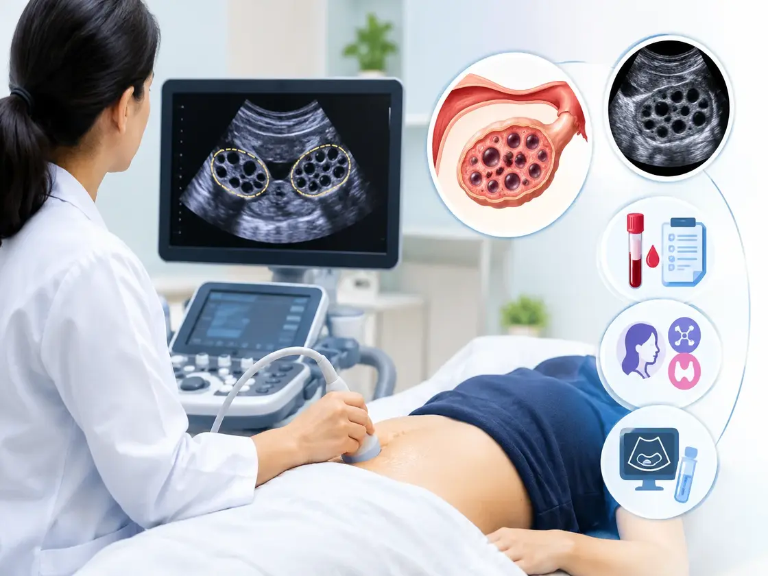
A pelvic ultrasound showing polycystic-appearing ovaries means your ovaries contain multiple small follicles, not that you definitely have PCOS. The scan is one piece of evidence, not the full diagnosis, which is why your doctor almost always pairs it with a hormone test in Indore or elsewhere before confirming anything. If you’ve just come out of a scan with unfamiliar terms on the report, here’s what they actually mean and what usually happens next.
What the Ultrasound Report Is Actually Describing
Your report will likely mention a few specific things about each ovary. Here’s what they mean in plain terms:
- Follicle count: The number of small, fluid-filled sacs visible on each ovary. A higher-than-typical count (often described as 12 or more per ovary in older criteria, or higher follicle density in newer ones) is one marker doctors look for.
- Ovarian volume: Enlarged ovaries are another common finding associated with PCOS.
- “String of pearls” appearance: a description sometimes used when follicles are arranged around the edge of the ovary, a visual pattern rather than a diagnosis on its own.
- Endometrial thickness: Doctors often check the lining of the uterus too, since irregular cycles common in PCOS can affect it.
None of these findings by themselves confirm PCOS. They’re descriptive, telling your doctor what the ovaries look like structurally, the same way a thyroid ultrasound describes a gland’s shape without saying how it’s functioning.
Why a Scan Alone Isn’t Enough
PCOS is officially diagnosed using a combination of factors, not the scan in isolation. Most doctors work from a framework requiring at least two of these three:
- Irregular or absent periods
- Signs of excess androgens (acne, excess hair growth, or blood test evidence of raised androgen levels)
- Polycystic-appearing ovaries on ultrasound
That third point is exactly why the scan alone can be misleading either way. Plenty of women without PCOS have ovaries that look polycystic on a scan, especially younger women, and plenty of women with PCOS have ovaries that look fairly ordinary. The report is context, not a verdict.
Confused about your ultrasound report? Bring it in and our radiologists can walk you through exactly what it means.
What Blood Tests Usually Follow
If your ultrasound raised the possibility of PCOS, the next step is almost always blood work to check hormone levels. A typical panel includes:
- A testosterone test in Indore, along with other androgens: Raised levels support a PCOS diagnosis
- FSH LH test in Indore: An elevated LH-to-FSH ratio is a pattern often seen in PCOS, though not always present. Many clinics group this with a broader fertility hormone test in Indore when fertility is also a concern
- AMH (Anti-Müllerian Hormone): Tends to run higher in women with PCOS and is increasingly used alongside ultrasound findings
- Fasting insulin and glucose: PCOS is closely linked to insulin resistance, so this is checked even without symptoms of diabetes
- Thyroid profile: Thyroid problems can cause similar cycle irregularities, so this is usually ruled out alongside everything else
This is the part where the ultrasound and the lab work start talking to each other. A scan finding on its own is a clue. A scan finding alongside raised androgens and an irregular cycle is a pattern doctors can actually act on.
Getting Both the Scan and the Blood Work Done Together
If you’re searching for sonography near me in Vijay Nagar, Indore after your doctor mentioned PCOS, the more useful question is usually whether the same place can also run the follow-up blood work, since ultrasound and hormone testing almost always go together here. Apoorva Diagnostic Centre runs both, an ultrasound scan in Indore covering pelvic and obstetric imaging, and a full hormone and pathology panel for androgens, LH, FSH, and related tests, across our Vijay Nagar and Sapna Sangeeta branches.
We’ve been trusted as one of the best diagnostic centre options in Indore for this exact combination, a scan and its follow-up blood work under one roof, for over 35 years, and we’re a pathology lab in Indore patients return to specifically for hormone panels like this one.
PCOS and Hair Fall, a Connected Symptom
Hair thinning is one of the more common signs that sends women looking into PCOS in the first place, alongside irregular periods and acne. If that’s part of what brought you here, our guide to hair fall in women covers how PCOS-related hair loss differs from thyroid or iron-related causes, and what to expect from that side of the workup.
Irregular periods, acne, and now an ultrasound flag? A hormone panel can complete the picture in one visit.
What Happens After a Confirmed PCOS Diagnosis
A diagnosis isn’t an endpoint, it’s the start of a management plan, and it looks different for everyone depending on what you’re trying to address, cycle regularity, fertility, or metabolic symptoms like insulin resistance. Your doctor typically decides next steps based on:
- Whether you’re trying to conceive or not
- How irregular your cycles are
- Whether blood work shows insulin resistance
- The degree of androgen-related symptoms
Ongoing monitoring often includes repeat ultrasounds and periodic hormone checks, particularly if medication or lifestyle changes are part of the plan, so your doctor can see whether things are moving in the right direction.
Frequently Asked Questions
1. Does a polycystic-looking ovary on ultrasound always mean I have PCOS?
No. Many women without PCOS have ovaries that appear polycystic on a scan, particularly at a younger age. Diagnosis requires additional criteria, usually irregular periods and evidence of raised androgens, alongside the scan.
2. What hormone tests are done to confirm PCOS?
Common tests include testosterone and other androgens, LH and FSH, AMH, and fasting insulin and glucose. A thyroid profile is often included too, since thyroid issues can cause similar symptoms.
3. Can PCOS be confirmed with ultrasound alone?
No. Ultrasound findings are one of three criteria doctors typically look for, alongside cycle irregularity and hormonal evidence. At least two of the three are usually needed for a diagnosis.
4. Is a transvaginal or abdominal ultrasound used to check for PCOS?
Both are used depending on the situation. A transvaginal scan generally gives a clearer view of ovarian structure, while an abdominal scan is used when transvaginal imaging isn’t appropriate or preferred.
5. How often should ultrasound and hormone tests be repeated after a PCOS diagnosis?
This depends on your treatment plan and symptoms, and your doctor will advise a schedule, often annually or alongside any medication changes, rather than a fixed universal timeline.
The Bottom Line
An ultrasound showing polycystic-appearing ovaries is a starting point, not a diagnosis by itself. What actually confirms PCOS is that finding combined with your cycle history and a hormone panel checking androgens, LH, FSH, and related markers. If your scan flagged something and you’re searching for a diagnostic centre near me in Indore that can handle the follow-up blood work too, getting it done at the same place as the scan usually means fewer appointments and a clearer picture, faster.
Been advised to have a hormone test or pelvic ultrasound? Call 0731 495 2233 to book at Apoorva Diagnostic Centre