
Ultrasound imaging is one of the most commonly used diagnostic tools in modern healthcare. It is safe, non-invasive, and provides valuable insights into different parts of the body. During pregnancy, ultrasound scans help doctors monitor the growth and development of the baby while ensuring the health of the mother.
Many patients often wonder about the difference between 2D, 3D, and 4D ultrasound and whether they need a specialized scan. While all three technologies use sound waves to create images, each offers a different level of detail and serves specific medical purposes.
At Apoorva Diagnostic Centre, we provide advanced ultrasound imaging services, including 2d 3d and 4d ultrasound in Indore, to help patients receive accurate and reliable diagnostic information.
Understanding the Difference Between 2D, 3D, and 4D Ultrasound
Before choosing a scan, it is important to understand how these ultrasound technologies work.
All ultrasound scans use high-frequency sound waves that bounce off tissues and organs. These reflected sound waves are converted into images by specialized equipment.
The main difference lies in how these images are processed and displayed.
What Is a 2D Ultrasound?
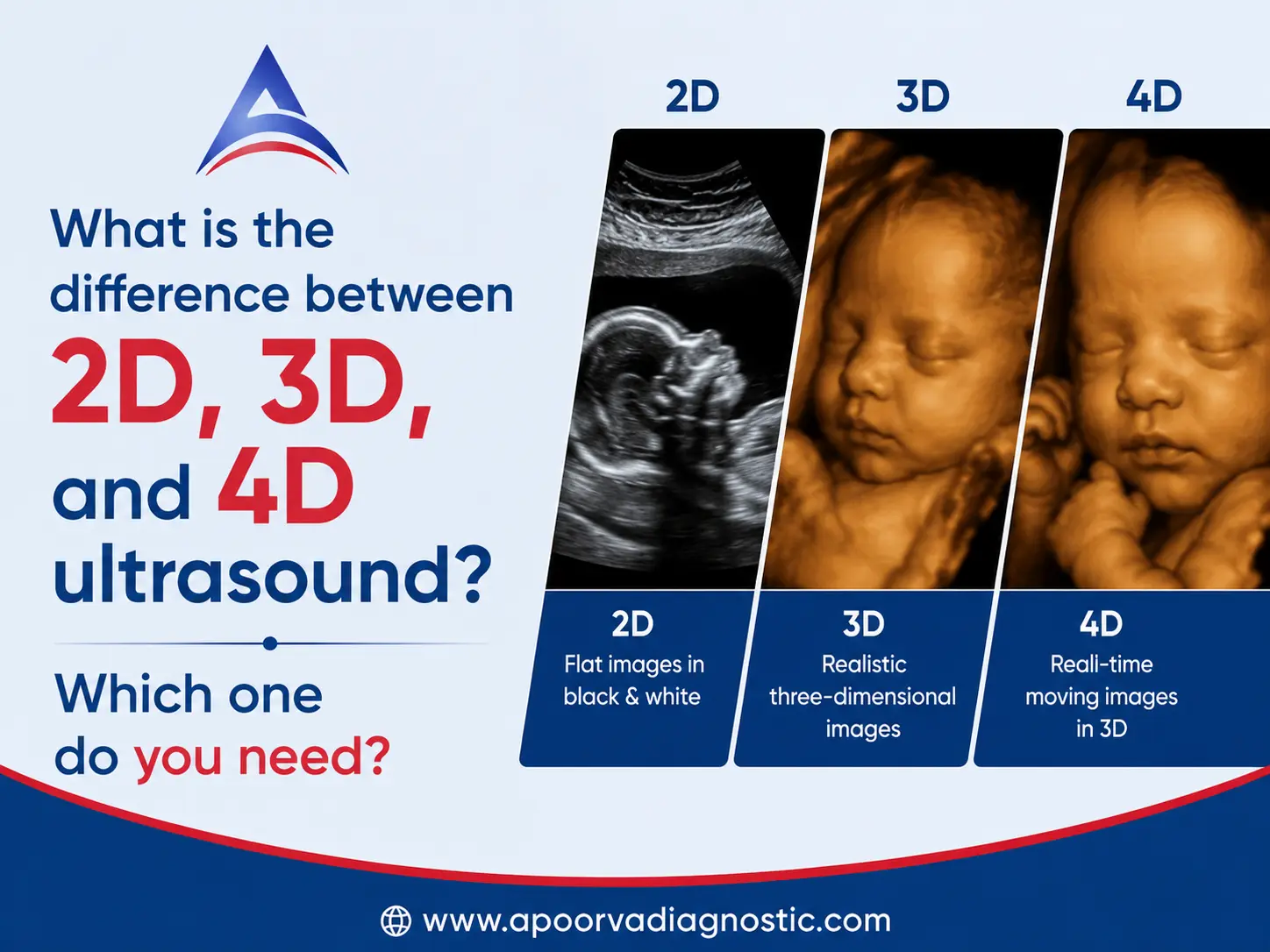
A 2D ultrasound is the traditional and most widely used form of sonography. It creates flat, black-and-white cross-sectional images of internal organs or a developing baby.
Doctors use 2D scans to:
- Confirm pregnancy
- Determine fetal age
- Monitor fetal growth
- Check organ health
- Detect abnormalities
- Evaluate blood flow when combined with Doppler imaging
2D ultrasound remains the standard imaging method because it provides clear diagnostic information and is highly effective for routine medical examinations.
What Is a 3D Ultrasound?
A 3D ultrasound combines multiple 2D images to create a three-dimensional image of the baby or internal organs.
Unlike traditional sonography, 3D scans provide depth, allowing doctors and parents to see facial features and body structures more clearly.
Benefits of 3D ultrasound include:
- Better visualization of fetal anatomy
- Enhanced detection of facial abnormalities
- Improved assessment of certain birth defects
- More detailed imaging of organs and tissues
Many expecting parents choose a 3d sonography for pregnancy in Indore because it provides a more realistic view of their unborn baby.
What Is a 4D Ultrasound?
A 4D ultrasound is essentially a live version of a 3D ultrasound. It produces real-time moving images.
Instead of a still image, parents can observe movements such as:
- Smiling
- Yawning
- Stretching
- Thumb sucking
- Hand movements
A 4d sonography in pregnancy offers a unique opportunity to observe fetal behavior while also helping doctors evaluate movement patterns.
The technology captures continuous 3D images over time, creating a live video effect.
Difference Between 2D, 3D, and 4D Ultrasound: A Detailed Comparison
| Feature | 2D Ultrasound | 3D Ultrasound | 4D Ultrasound |
| Image Type | Flat image | Three-dimensional image | Live moving 3D image |
| Color | Black and white | Detailed surface image | Real-time detailed image |
| Diagnostic Use | Routine examinations | Detailed anatomical assessment | Movement evaluation |
| Pregnancy Monitoring | Excellent | Excellent | Excellent |
| Visualization of Baby | Basic | Advanced | Most realistic |
| Cost | Lowest | Moderate | Higher |
| Availability | Widely available | Specialized centres | Advanced centres |
This comparison clearly highlights the difference between 2D, 3D, and 4D ultrasound and helps patients choose the most suitable option.
Why Is 2D Ultrasound Still the Most Common Choice?
Many patients assume newer technologies automatically replace older ones. However, this is not the case.
2D ultrasound remains the primary imaging tool because it:
- Provides excellent diagnostic accuracy
- Is cost-effective
- Supports routine pregnancy monitoring
- Detects most fetal abnormalities
- Is recommended for standard prenatal care
Doctors continue to rely heavily on 2D ultrasound for medical decision-making.
Benefits of 3D Sonography for Pregnancy
Many pregnant women opt for 3d sonography for pregnancy in Indore because it offers enhanced visualization.
Some benefits include:
Better Facial Imaging
Parents can see facial features more clearly, creating a memorable bonding experience.
Improved Detection of Structural Issues
3D imaging may help doctors evaluate:
- Cleft lip
- Facial abnormalities
- Skeletal conditions
- Neural tube defects
Enhanced Patient Understanding
Three-dimensional images often make it easier for parents to understand fetal development.
Benefits of 4D Sonography in Pregnancy
A 4D sonography in pregnancy provides live imaging of fetal movements.
Advantages include:
Real-Time Observation
Parents can watch the baby move naturally inside the womb.
Evaluation of Movement Patterns
Doctors can assess:
- Limb movement
- Facial expressions
- Behavioral development
Emotional Bonding
Many families find 4D imaging an exciting and memorable experience during pregnancy.
When Is a 3D or 4D Ultrasound Recommended?
Although routine pregnancy monitoring is usually performed using a 2D ultrasound, doctors may recommend advanced imaging in specific situations.
These include:
- Suspected fetal abnormalities
- High-risk pregnancies
- Detailed anatomical assessment
- Monitoring specific developmental concerns
- Enhanced visualization requirements
A qualified radiologist or sonologist will determine whether additional imaging is medically necessary.
Is 4D Ultrasonography Safe During Pregnancy?
Safety is one of the most common concerns among expecting parents.
Current medical evidence indicates that ultrasound procedures, including 4d ultrasonography test in Indore, are considered safe when performed by trained healthcare professionals and only when medically appropriate.
Ultrasound uses sound waves rather than ionizing radiation, making it safer than many imaging techniques.
However, healthcare providers recommend avoiding unnecessary scans and following medical guidelines.
Best Time for a 3D or 4D Pregnancy Scan
Timing plays an important role in obtaining clear images.
Most specialists recommend scheduling a 3D or 4D scan between:
26 to 32 Weeks of Pregnancy
During this period:
- Facial features are well developed.
- The baby has sufficient fat beneath the skin.
- Amniotic fluid levels often support better image quality.
This timeframe usually provides the best results for 3d 4d sonography test in Indore.
Who Should Consider 2D, 3D, or 4D Ultrasound?
Different patients have different needs.
Choose 2D Ultrasound If:
- You need routine pregnancy monitoring.
- Your doctor requests standard diagnostic imaging.
- You want a cost-effective examination.
Choose 3D Ultrasound If:
- Detailed anatomical evaluation is needed.
- Your doctor wants enhanced visualization.
- You wish to see clearer fetal images.
Choose 4D Ultrasound If:
- You want real-time fetal movement visualization.
- Your doctor recommends advanced fetal assessment.
- You seek a more engaging pregnancy experience.
Understanding the difference between 2D, 3D, and 4D ultrasound can help you make an informed decision with your healthcare provider.
Why Choose Apoorva Diagnostic Centre for Ultrasound Services?
At Apoorva Diagnostic Centre, we are committed to delivering accurate imaging services using advanced diagnostic technology.
Our advantages include:
- Experienced radiologists
- Modern ultrasound equipment
- Comfortable patient environment
- Accurate and timely reports
- Affordable imaging services
- Comprehensive pregnancy scans
Whether you need a routine pregnancy scan or advanced 2d 3d and 4d ultrasound in Indore, our team is dedicated to providing reliable diagnostic support.
Frequently Asked Questions
1. What is the main difference between 2D, 3D, and 4D ultrasound?
The primary difference is how images are displayed. 2D ultrasound produces flat images, 3D ultrasound creates detailed three-dimensional images, and 4D ultrasound shows live moving 3D images in real time.
2. Is a 4D ultrasound better than a 2D ultrasound?
Not necessarily. A 2D ultrasound remains the standard diagnostic tool for most medical evaluations. A 4D ultrasound offers enhanced visualization and live imaging but is often used as a complementary examination.
3. When should I get a 3D or 4D pregnancy scan?
Most specialists recommend scheduling a 3D or 4D scan between 26 and 32 weeks of pregnancy for optimal image quality and fetal visibility.
Book Your Ultrasound Appointment Today
Looking for reliable 2d 3d and 4d ultrasound in Indore? Apoorva Diagnostic Centre offers advanced imaging services with experienced specialists and modern diagnostic technology.
Whether you require a routine pregnancy scan, 3d sonography for pregnancy in Indore, or a detailed 4d ultrasonography test in Indore, our team is here to help.
Contact Apoorva Diagnostic Centre today to schedule your ultrasound appointment and receive accurate diagnostic care for you and your family.